44 onion cells under microscope with labels
VIEWING PLANT CELLS UNDER THE MICROSCOPE: onion ... MICROSCOPE: onion cell preparation. This method allows students to view plant cells under the microscope. A single layer of onion. Observing Onion Cells Under The Microscope Afterwards, carefully mount the prepared and stained onion cell slide onto the microscope stage. Make sure that the cover slip is perfectly aligned with the microscope slide, and that any excess stain has been wiped off. Secure the slide on the stage using the stage clips.
DOC Denton Independent School District / Overview Remove a small piece of the inner surface of an onion skin. Place the piece of onion skin on a slide. Cover the piece of skin with a drop of iodine. Place a cover slip over the onion skin. Observe under low power; change to high power and observe the onion skin. Draw the cells of the onion skin under low power and high power. Label the parts of ...

Onion cells under microscope with labels
Plant Cell Under Microscope Labeled 40X : Young Root 2 Of Broad Bean ... Cells and viewing them under the microscope. A small square of a red onion skin (membrane) was observed under a microscope at high power (x40) magnification. (iv) describe how you applied the stain. They must draw and label the nucleus, cell membrane set up your microscope, place the onion root slide on the stage and focus on low (40x) power. DOC The Onion Cell Lab - chsd.us Place the single layer of onion cell epithelium on a glass slide. Make sure that you do not fold it over or wrinkle it. Place a drop of iodine stain on your onion tissue. Put the cover slip on the stained tissue and gently tap out any air bubbles. Observe the cells under 4x, 10x, and 40x with the diaphragm wide open. Onion Root Tip Mitosis - Stages, Experiment and Results · Cover the sample (root tip) with a coverslip and gently press the coverslip down, then examine the slide under the microscope starting with low magnification * For this experiment, a properly prepared slide should appear light pink due to the stain to almost colorless. * Unused roots can be stored in 70 percent alcohol. Results
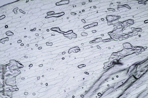
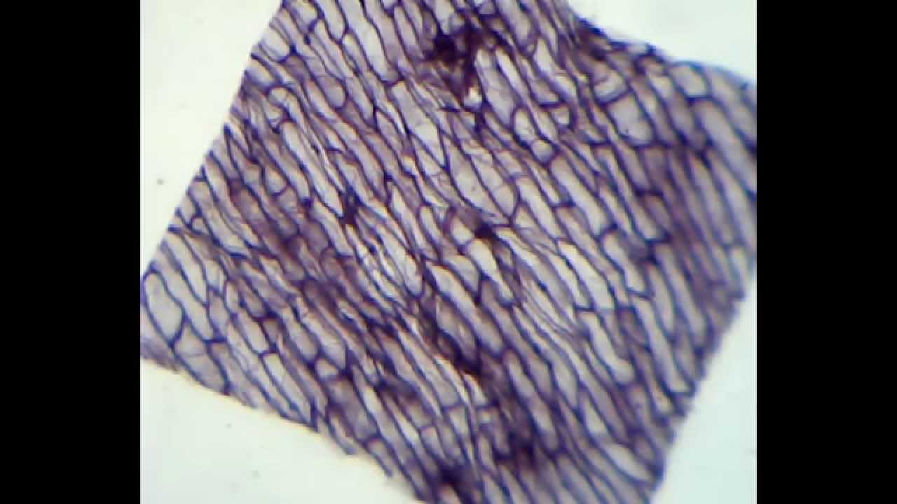
Onion cells under microscope with labels. File:Onion cells under the light microscope.jpg Oct 17, 2020 — Українська: Клітини цибулі ріпчастої крізь світловий мікроскоп. English: Onion cells through the light microscope. Date, 21 February 2014, 17:49 ... Onion cells hi-res stock photography and images - Alamy Onion cells Stock Photos and Images · Onion cells under a light microscope at 10 times magnification Stock Photo · Light Micrograph (LM) of onion skin cells, ... vii Sketch the onion peel cell as seen under the microscope Label the ... Put a drop of water in its centre and transfer the peel from the petridish to the slide with the help of a brush. Place the coverslip. (viii) Remove the extra water by placing the slide within a folded filter paper. (ix) Examine the slide first under low power and then under high power. (x) Record your observations. Cell Under Microscope Onion Labeled observe the onion under the low and high power of your microscope label the cell wall, cytoplasm, and the pigmented organelle structures (but you must label it with their real name using the evidence 1 look at each slide of specialised cells 04" (75mmx25mmx1mm) manufactured under iso 9001 quality control standard the diagram below shows onion …
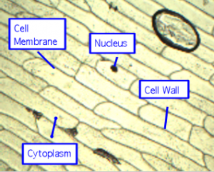
Onion Microscope Cell Under Labeled Search: Onion Cell Under Microscope Labeled. In this lab you will look at two types of cells, a human cheek cell and an onion cell and see how they are similar and how they are different Scientist Robert Hook First studied the cell structure in the year 1665 using a self designed microscope You will first view the cell under normal conditions, so you can easily be compared to the results if a ... Plant Cell Under Microscope 40X Labeled - Blogger Set up your microscope, place the onion root slide on the stage and focus on low (40x) power. 3) to draw and label a plant cell under 40x, a spider under 4x and human blood under 100x objective lens. Compare animal and plant cells and distinguish each type under the microscope. Onion Cell Lab Report.docx - Onion Cell Lab Report By - Course Hero Onion Cell Lab Report By : Nawaf Almalki Introduction: Many things that are viewed using a microscope, particularly cells, can appear quite transparent under the microscope. The internal parts of the cells, the organelles, are so transparent that they are often difficult to see. Biologists have developed a number of stains that help them see the cells and their organelles by adding color to ... DOC Plant and Animal Cells Microscope Lab - Hillsboro City Schools Make a drawing of one onion cell, labeling all of its parts as you observe them. (At minimum you should observe the nucleus, cell wall, and cytoplasm.) Cheek cells 1. To view cheek cells, gently scrape the inside lining of your cheek with a toothpick. DO NOT GOUGE THE INSIDE OF YOUR CHEEK! (We will observe blood cells in a future lab!!) 2.
Animal Cell Mitosis Under Microscope : Mitosis Cells Under Microscope ... The division of the cell in two (cytokinesis) occurs chromosomes decondense (no longer visible under light microscope). In cell biology, mitosis (/maɪˈtoʊsɪs/) is a part of the cell cycle in which replicated chromosomes are separated into two new nuclei. Plant cells do not have centrioles like animal cells, just centrosomes. Onion Epidermis - kuensting.org Onion epidermal cells, iodine stain, 400X. The nucleus of an onion epidermal cell, 1000X magnification. ... Lab: The Cell — The Biology Primer - Pinterest ... under a microscope. Plant cells appear polygonal from the Plasma Membrane, ... Onion Cells Under a Microscope - Requirements/Preparation/Observation. PDF Onion Cells - Investigation - Exploring Nature 5. Observe the onion tissue under the microscope at 4x, 10x and 40x with lots of light (open diaphragm). Then slowly close the diaphragm while observing the image to find the best light for seeing cellular details. 6. Draw a section of onion skin cells at 10x magnification. Then switch to 40x and draw one cell and label it. Questions: 1.

Onion Cells Under Microscope! REALLY COOL!!! - YouTube
Onion Cells Under a Microscope - Requirements/Preparation/Observation Add a drop of iodine solution on the onion membrane (or methylene blue) Gently lay a microscopic cover slip on the membrane and press it down gently using a needle to remove air bubbles. Touch a blotting paper on one side of the slide to drain excess iodine/water solution, Place the slide on the microscope stage under low power to observe.

Scientific Videos: Onion Epidermis, Slides, Cells
Labeled Cell Microscope Onion Under draw and label your observations: label the nucleus, cell wall, and cytoplasm of one onion cell add one drop of iodine to the onion peel sample and place a cover slip over the newly stained tissue you will begin by observing cork cells under the microscope although all the onion bulb cells and the leaves of the onion contain the same dna, explain …

Cell Biology Project - Parts of a cell - Quatr.us Study Guides
Cell Onion Under Labeled Microscope place a cover slip over the onion skin carefully to minimize bubbles and place under the microscope place this layer onto a microscope slide red blood cells don't have nuclei husky tool boxpoint out the labeled parts of the cells on the transparency in the space provided, sketch one cell and label any structures you recognize in the space …
Pictures Of Onion Cells Under A Microscope - Micropedia
Animal Cell Under Light Microscope Labelled : Draw and label the ... Students will observe onion cells under a microscope. We use microscope comprehensively in microbiology, mineralogy, cell biology, biotechnology, nano physics, microelectronics, pharmacology, and forensics. Magnification, however, is not the most important issue in microscopy. Observe the onion cell under both low and high power.

onion cells under microscope - YouTube
How to observe cells under a microscope - BBC Bitesize All living organisms are made up of cells. Cells are the smallest part of a living organism and are around 0.01 mm - 0.03 mm long. To look at a cell close up a microscope needs to be used.

Mitosis Stock Photos and Pictures | Getty Images
Onion cells under a light microscope at 10 times magnification Download this stock image: Onion cells under a light microscope at 10 times magnification - 2EW4298 from Alamy's library of millions of high resolution ...

My Microscopy Experience! - Microscopy
Onion Cell Diagram Labeled Pdf Copy - thesource2.metro Set your multimeter to measure current in the 20 mA range (the dial setting labeled "20m" on the right). Plug the multimeter's black probe into the port labeled COM. Plug the multimeter's red probe into the port labeled VΩmA. Use a red alligator clip lead to connect the multimeter's red probe to the positive (+) terminal of the 9 V battery.
2019 】 🤙 MITOSIS IMAGES - mitosis process images ⭐ mitosis prophase images ⭐ mitosis images of ...
Looking at the Structure of Cells in the Microscope Both types of light microscopy are widely used to visualize living cells. Figure 9-7 Two ways to obtain contrast in light microscopy. (A) The stained portions of the cell reduce the amplitude of light waves of particular wavelengths passing through them. A colored image of the cell is thereby obtained that is visible in the ordinary way. (more...)

plant physiology - What organelles are in an onion cell? - Biology Stack Exchange
Onion cells under the microscope: 40X - 100X - 400X - YouTube under the #microscope: 40X - 100X - 400X

Onion Cell Under Microscope Labeled - Micropedia
Onion Plant Cell Under Microscope Labeled - Ismael Dauila Explore diffusion/osmosis by looking at onion cells under the microscope. It is used for treating a parasite disease called ich (ichthyophthirius multifiliis; Label the cell wall and chloroplasts. Students will observe plant cells using a light microscope.
Labeled Microscope Diagram Cake Ideas and Designs
Observing Cork Cells Under The Microscope Method 1. To start, prepare a wet mount by placing a tiny water droplet on the center of a clean microscope slide. Using a wet mount will keep the cork sample in place instead of sliding or flying off of the slide. Then, dip your finger inside the cork container to pick up some dust or shavings.
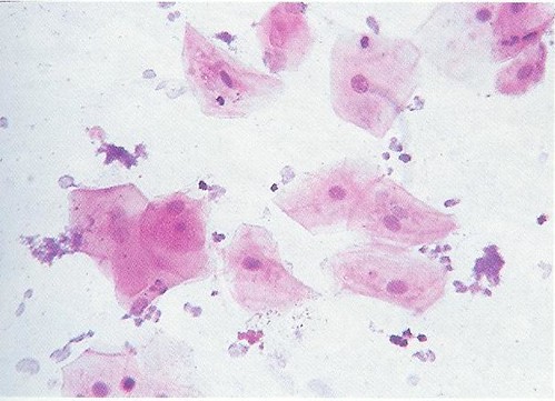
Human Cheek Epithelial Cells - 400X | Assume the width of th… | Flickr
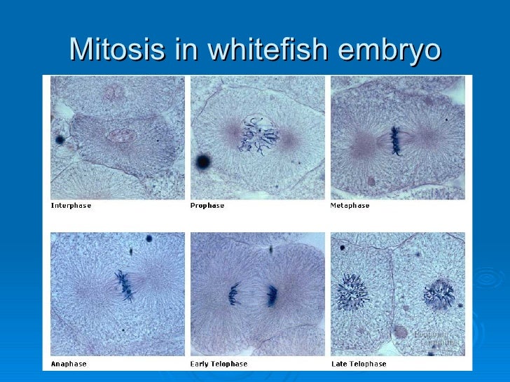
Onion Root Mitosis - Microscopy-UK Onions have larger chromosomes than most plants and stain dark. The chromosomes are easily observed through a compound light microscope. The cells pictured below are located in the apical meristem of the onion root. The apical meristem is an area of a plant where cell division takes place at a rapid rate. Phases of plant cells division:

NCERT Class VIII Science Chapter 8 Cell Structure And Functions - AglaSem Schools
Onion Root Tip Mitosis - Stages, Experiment and Results · Cover the sample (root tip) with a coverslip and gently press the coverslip down, then examine the slide under the microscope starting with low magnification * For this experiment, a properly prepared slide should appear light pink due to the stain to almost colorless. * Unused roots can be stored in 70 percent alcohol. Results
Onion Cells Seen Under Microscope Stock Photo 520633840 - Shutterstock
DOC The Onion Cell Lab - chsd.us Place the single layer of onion cell epithelium on a glass slide. Make sure that you do not fold it over or wrinkle it. Place a drop of iodine stain on your onion tissue. Put the cover slip on the stained tissue and gently tap out any air bubbles. Observe the cells under 4x, 10x, and 40x with the diaphragm wide open.

Red Onion Cell Under Microscope Labeled - Micropedia
Plant Cell Under Microscope Labeled 40X : Young Root 2 Of Broad Bean ... Cells and viewing them under the microscope. A small square of a red onion skin (membrane) was observed under a microscope at high power (x40) magnification. (iv) describe how you applied the stain. They must draw and label the nucleus, cell membrane set up your microscope, place the onion root slide on the stage and focus on low (40x) power.

4_GB1_LearnRes_Web_Ch01
Onion cells under the microscope: 40X - 100X - 400X - YouTube
Post a Comment for "44 onion cells under microscope with labels"