41 human eye with labels
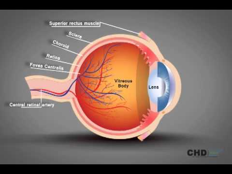
Human Eye: Structure of Human Eye (With Diagram) | Biology The human eye is a very sensitive and delicate organ suspended in the eye socket which protects it from injuries. It essentially consists of CORNEA, LENS & RETINA besides many other parts such as Iris, Pupil and aqueous humour, vituous humour etc. Each one has got a specific function. A section of the eye is as shown in Fig. 2.2. Anatomy of the eye: Quizzes and diagrams | Kenhub How to learn the parts of the eye. Found within two cavities in the skull known as the orbits, the eyes are surrounded by several supporting structures including muscles, vessels, and nerves.There are 7 bones of the orbit, two groups of muscles (intrinsic ocular and extraocular), three layers to the eyeball… and that's just the beginning. There's a lot to learn, but stay calm!
How to draw the Human Eye - Labeled Science Diagrams - YouTube Download a free printable outline of this video and draw along with us: you for watching. Please subsc...

Human eye with labels
6,819 Human eye diagram Images, Stock Photos & Vectors - Shutterstock 6,819 human eye diagram stock photos, vectors, and illustrations are available royalty-free. See human eye diagram stock video clips Image type Orientation Color People Artists Sort by Popular Biology Healthcare and Medical Icons and Graphics Diseases, Viruses, and Disorders human eye anatomy 3d rendering eye medicine retina Next of 69 Labeled Eye Diagram | Science Trends What you want to interpret as a major part of the human eye is somewhat up to the individual, but in general there are seven parts of the human eye: the cornea, the pupil, the iris, the lens, the vitreous humor, the retina, and the sclera. Let's take a closer look at each of these components individually. The Cornea Label Parts of the Human Eye - University of Dayton Parts of the Eye. Select the correct label for each part of the eye. The image is taken from above the left eye. Click on the Score button to see how you did. Incorrect answers will be marked in red. ...
Human eye with labels. The Eyes (Human Anatomy): Diagram, Optic Nerve, Iris, Cornea ... - WebMD The front part (what you see in the mirror) includes: Iris: the colored part. Cornea: a clear dome over the iris. Pupil: the black circular opening in the iris that lets light in. Sclera: the ... Eye Anatomy: 16 Parts of the Eye & Their Functions - Vision Center The following are parts of the human eyes and their functions: 1. Conjunctiva The conjunctiva is the membrane covering the sclera (white portion of your eye). The conjunctiva also covers the interior of your eyelids. Conjunctivitis, often known as pink eye, occurs when this thin membrane becomes inflamed or swollen. Structure and Functions of Human Eye with labelled Diagram - BYJUS The human eye is a roughly spherical organ, responsible for perceiving visual stimuli. It is enclosed within the eye sockets in the skull and is anchored down by muscles within the sockets. Anatomically, the eye comprises two components fused into one; hence, it does not possess a perfect spherical shape. FREE! - The Human Eye Labeling Activity (Teacher-Made) - Twinkl In this resource, you'll find a 2-page PDF that is easy to download, print out, and use immediately with your class. The first page is a labelling exercise with two diagrams of the human eye. One is a view from the outside, and the other is a more detailed cross-section. Challenge learners to label the parts of the eye diagram. On the second page, you'll find a set of answers showing ...
Labelling the eye — Science Learning Hub Labelling the eye Resource Add to collection The human eye contains structures that allow it to perceive light, movement and colour differences. In this activity, students use online or paper resources to identity and label the main parts of the human eye. By the end of this activity, students should be able to: PDF Parts of the Eye - National Institutes of Health Eye Diagram Handout Author: National Eye Health Education Program of the National Eye Institute, National Institutes of Health Subject: Handout illustrating parts of the eye Keywords: parts of the eye, eye diagram, vitreous gel, iris, cornea, pupil, lens, optic nerve, macula, retina Created Date: 12/16/2011 12:39:09 PM Labelled Diagram of Human Eye, Explanation and Function - VEDANTU The human eye is a part of the sensory nervous system. Labeled Diagram of Human Eye The eyes of all mammals consist of a non-image-forming photosensitive ganglion within the retina which receives light, adjusts the dimensions of the pupil, regulates the availability of melatonin hormones, and also entertains the body clock. Human Eye Anatomy Pictures, Images and Stock Photos Medical icons of internal human organs realised in modern flat design with long shadow. Eye layers The inner layer of the eye, or retina, is similar to film in a camera. It receives light from an image we are looking at, and converts that light into electrical impulses which are sent through the fibers of the optic nerve to the brain.
Human eye diagram to label - simplediagram.netlify.app Human eye diagram labeled parts of the human eye diagram and human eye diagram labeled are three main things we want to present to you based on the post title. Layer of cells on the back of the eye. Controls how much light enters the eye. To play the game online visit Labeling Parts of the Eye 5th Grade. In the 2nd worksheet they match the. 60,892 Human eye anatomy Images, Stock Photos & Vectors - Shutterstock Find Human eye anatomy stock images in HD and millions of other royalty-free stock photos, illustrations and vectors in the Shutterstock collection. Thousands of new, high-quality pictures added every day. Eye Diagram With Labels and detailed description - BYJUS A brief description of the eye along with a well-labelled diagram is given below for reference. Well-Labelled Diagram of Eye The anterior chamber of the eye is the space between the cornea and the iris and is filled with a lubricating fluid, aqueous humour. The vascular layer of the eye, known as the choroid contains the connective tissue. Eye Anatomy: A Closer Look At the Parts of the Eye - All About Vision In a number of ways, the human eye works much like a digital camera: Light is focused primarily by the cornea — the clear front surface of the eye, which acts like a camera lens. The iris of the eye functions like the diaphragm of a camera, controlling the amount of light reaching the back of the eye by automatically adjusting the size of the ...
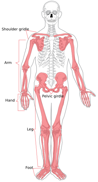
Appendicular Skeleton Diagram Clip Art at Clker.com - vector clip art online, royalty free ...
HOW TO DRAW HUMAN EYE WITH LABELLING - YouTube Let's learn HOW TO DRAW HUMAN EYE WITH LABELLING. Follow my drawing of human eyes step by step and I am sure you will draw it easily. This is a suitable draw...

Human Anatomy Lab: The Urinary and Reproductive Systems
Anatomy of the Eye | Johns Hopkins Medicine Ciliary body. The part of the eye that produces aqueous humor. Cornea. The clear, dome-shaped surface that covers the front of the eye. Iris. The colored part of the eye. The iris is partly responsible for regulating the amount of light permitted to enter the eye. Lens (also called crystalline lens).
Human Eye Structure: Eye Anatomy Explained - YouTube
The Human Eye (Eyeball) Diagram, Parts and Pictures The human eye consists of the eyeball, optic nerve, orbit and appendages (eyelids, extraocular muscles and lacrimal glands). While the eyeball is the actual sensory organ, the other parts of of the eye are equally important in maintaining the health and function of the eye as a whole. The structure of the human eye is such that light can enter ...

Human Skeleton Blank Clip Art at Clker.com - vector clip art online, royalty free & public domain
Human eye model labeled Flashcards | Quizlet all of the open space. Lacrimal Gland. Fovea centralis. the black dot in the middle. Medial Commissure. Lacrimal Ducts. Macula Lutea. The pink dot (not the black in the middle)

Common Types of Warehouse Labels | ID Label Inc.
Labelling the eye — Science Learning Hub In this interactive, you can label parts of the human eye. Use your mouse or finger to hover over a box to highlight the part to be named. Drag and drop the text labels onto the boxes next to the eye diagram If you want to redo an answer, click on the box and the answer will go back to the top so you can move it to another box.

Human Anatomy Lab: Muscles of the Leg
Human eye - Wikipedia The human eye is a sensory organ, part of the sensory nervous system, that reacts to visible light and allows us to use visual information for various purposes including seeing things, keeping our balance, and maintaining circadian rhythm . The eye can be considered as a living optical device.

Beyond the Human Eye: A tiny aquatic worm that clones itself
Human Eye Diagram, How The Eye Work -15 Amazing Facts of Eye Fun Facts About Human Eye For Kids FACT 1 Iris scanning is more secure than fingerprints because our iris has 256 unique characteristics and the fingerprint has just 40. FACT 2 Newborn babies don't produce tears. They only make crying sounds, but no tears come out of their crying eyes.

I Have Seen The Whole Of The Internet: Woman With Blood Vessel In Her Eye That Spells Love
Structure of the Human Eye - Health Jade The eye is a hollow, spherical structure about 2.5 centimeters in diameter. Its wall has three distinct layers—an outer (fibrous) layer, a middle (vascular) layer, and an inner (nervous) layer. The spaces within the eye are filled with fluids that help maintain its shape. Figure 6. Structure of the human eye.
Post a Comment for "41 human eye with labels"