43 light microscope with labels
Light Microscope-Definition, Principle, Types, Parts, Labeled Diagram ... A light microscope is a device or instrument used in biology laboratories that uses visible light to locate, magnify, and expand micro objects. Using lenses, they focus light on the specimen and magnify it to generate a photograph. Typically, the specimen is positioned close to the microscopic lens. A practical guide to scanning light-field microscopy with digital ... Set the number of scanning light-field images you want to capture in the ' Image Num ' box. Close the live mode by clicking the ' CloseCam ' button. 31. Open data-saving mode with real ...
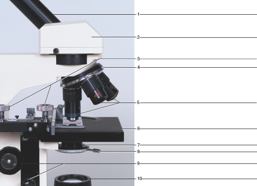
An Introduction to the Light Microscope, Light Microscopy Techniques ... a) standard upright microscope indicating (1) e yepiece (ocular lens), (2) objective turret, revolver, or revolving nose piece (to hold multiple objective lenses), (3) objective lenses, focus knobs (to move the stage) (4) coarse adjustment, (5) fine adjustment, (6) stage (to hold the specimen), (7) light source (a light or a mirror), (8) …
Light microscope with labels
Compound Microscope- Definition, Labeled Diagram, Principle, Parts, Uses The common light microscope is also called a bright-field microscope because the image is produced amidst a brightly illuminated field. The image appears darker because the specimen or object is denser and somewhat opaque than the surroundings. Part of the light passing through or object is absorbed. Magnification of compound microscope ZEISS Lattice Lightsheet 7 Your automated and easy-to-use lattice light-sheet microscope for long-term volumetric imaging of living cells at subcellular resolution. ... Time lapse movie showing dynamics of a U2OS cell stably expressing Actin-GFP (cytoskeleton, cyan). Cells were also labeled with MitoTracker™ Red CMXRos (Mitochondria, green) and Draq 5 (Nucleus, magenta Light Microscope- Definition, Principle, Types, Parts, Labeled Diagram ... A light microscope is a biology laboratory instrument or tool, that uses visible light to detect and magnify very small objects and enlarge them. They use lenses to focus light on the specimen, magnifying it thus producing an image. The specimen is normally placed close to the microscopic lens.
Light microscope with labels. Light-sheet microscopy at high resolution - Nature An improved light-sheet microscope images live cells at sub-100-nm axial resolution. New light microscopy techniques designed to improve spatial resolution are often less widely applicable than ... Dissecting microscope (Stereo or stereoscopic microscope)- Definition ... Figure: Labeled Dissecting microscope (Stereo or stereoscopic microscope). Image created using biorender.com. LED illuminators-For some of the dissecting Microscopes, they have an inbuilt LED illuminator as a source of light.Eyepieces-They have two eyepieces each focusing different pathways of the light into and out of the specimen, each with its own magnification power. Researchers demonstrate label-free super-resolution microscopy A newly developed sub-diffraction-limit microscopy approach doesn't require fluorescent labels. The video shows the process of the data evaluation algorithm, retrieving the positions and sizes of... Brightfield Microscope (Compound Light Microscope)- Definition ... Brightfield Microscope Definition Brightfield Microscope is also known as the Compound Light Microscope. It is an optical microscope that uses light rays to produce a dark image against a bright background. It is the standard microscope that is used in Biology, Cellular Biology, and Microbiological Laboratory studies.
Neuron under Microscope with Labeled Diagram - AnatomyLearner Neuron under Microscope with Labeled Diagram. 31/03/2022 31/03/2022 by anatomylearner. The structural and functional unit of the nervous system is the neuron that may easily observe under a light microscope. Neurons may vary considerably in size, shape, and other features. Here, I will provide details information and identifying points so that ... Bright-field microscope (Compound light microscope) - Diagram (Parts ... Bright-field microscope parts (Labeled Diagram) Ocular Lens This microscope has two eye lenses or ocular lens on the top of the microscope that are used to focus the image from the objective lens. It is from these lenses that we see the magnified image of the specimen. Objective Lens Compound Microscope - Diagram (Parts labelled), Principle and Uses Compound Microscope - Diagram (Parts labelled), Principle and Uses As the name suggests, a compound microscope uses a combination of lenses coupled with an artificial light source to magnify an object at various zoom levels to study the object. A compound microscope: Is used to view samples that are not visible to the naked eye Microscope Types (with labeled diagrams) and Functions This is an advanced microscope that has specific application in viewing, observing and measuring the optical thickness and phase of completely transparent specimens and objects. A tiny interferometer is used and a specimen is placed on beam path of it. This path is split and then rejoined to create two superimposed images of the specimen in focus.
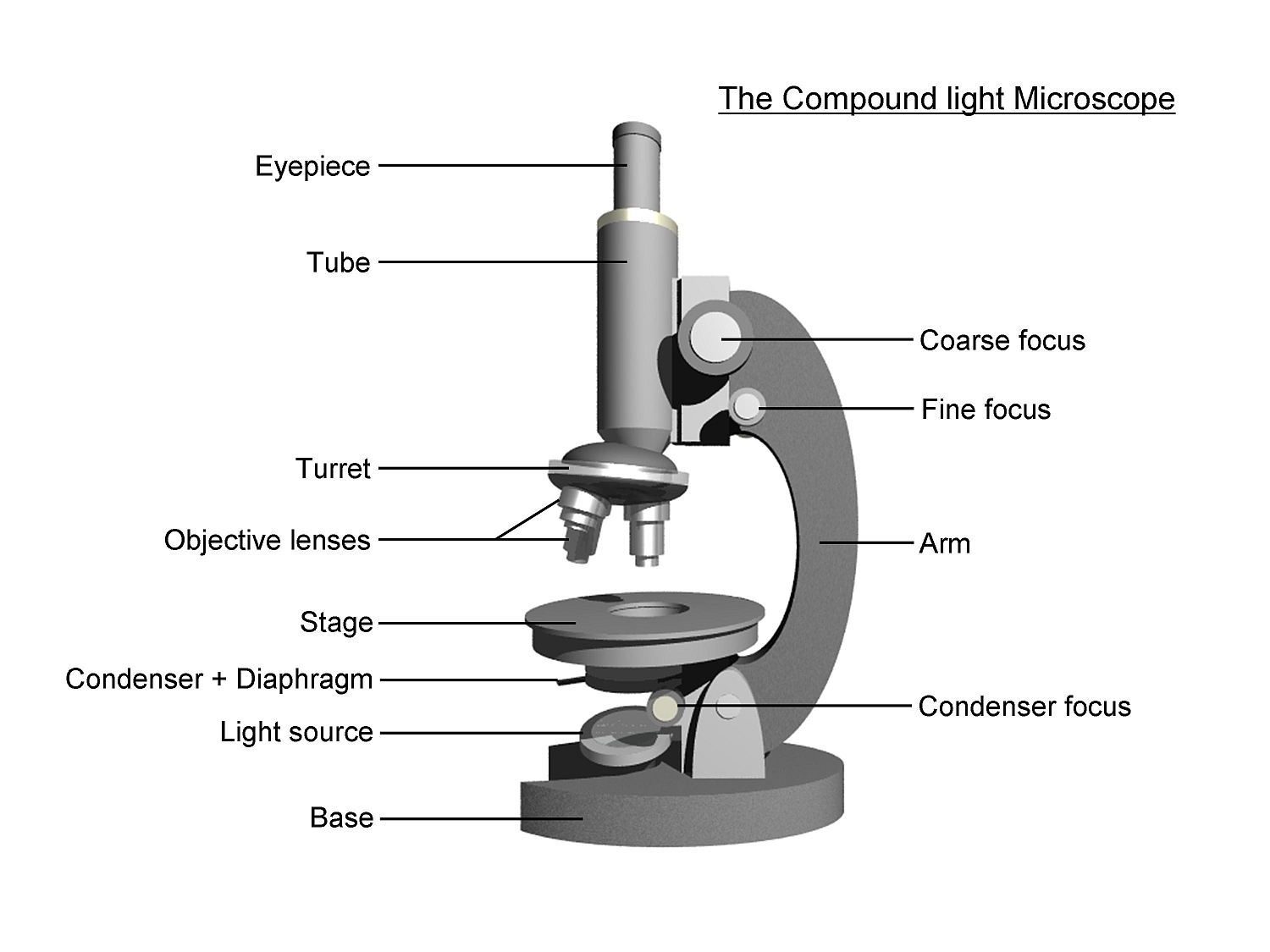
Microscope Parts, Function, & Labeled Diagram - slidingmotion Microscope parts labeled diagram gives us all the information about its parts and their position in the microscope. Microscope Parts Labeled Diagram The principle of the Microscope gives you an exact reason to use it. It works on the 3 principles. Magnification Resolving Power Numerical Aperture. Parts of Microscope Head Base Arm Eyepiece Lens 1.5: Microscopy - Biology LibreTexts In Biology, the compound light microscope is a useful tool for studying small specimens that are not visible to the naked eye. The microscope uses bright light to illuminate through the specimen and provides an inverted image at high magnification and resolution. ... Blank microscope to label parts. 1.5: Microscopy is shared under a CC BY 4.0 ... Simple Microscope - Diagram (Parts labelled), Principle, Formula and Uses A simple microscope consists of Optical parts Mechanical parts Labeled Diagram of simple microscope parts Optical parts The optical parts of a simple microscope include Lens Mirror Eyepiece Lens A simple microscope uses biconvex lens to magnify the image of a specimen under focus. Light Microscope Labeled Gcse - 17 images - how are concave and convex ... Light Microscope Labeled Gcse. Here are a number of highest rated Light Microscope Labeled Gcse pictures upon internet. We identified it from reliable source. Its submitted by doling out in the best field. We acknowledge this kind of Light Microscope Labeled Gcse graphic could possibly be the most trending topic later than we allocation it in ...
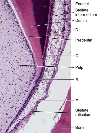
7: Enamel: Composition, Formation, and Structure | Pocket Dentistry
Types and parts of microscopes - Kenhub Light microscopy is probably the most popular form of microscopy encountered by students. It uses light from the visible spectrum and couples it with compound (serial) magnifying lenses to observe an object. Light Microscope There are several components to the modern light microscope that come together to enhance its function:

Foram, Foraminifera, 200 X, Protozoa, optical microscope Stock Photo - Alamy
Simple Microscope - Parts, Functions, Diagram and Labelling Confocal microscope - It uses laser light to scan a dyed sample. Scanning electron microscope - Instead of light, this type of microscope uses electron. This type of microscope is used by researchers in the field of physical, biological, and medical science. Transmission electron microscope - it uses electron to create a magnified image.
Label the Light Microscope - Labelled diagram
Parts of a microscope with functions and labeled diagram Microscopic illuminator - This is the microscopes light source, located at the base. It is used instead of a mirror. It captures light from an external source of a low voltage of about 100v. Condenser - These are lenses that are used to collect and focus light from the illuminator into the specimen.
32 Compound Light Microscope Label - Labels 2021
Pancreas Histology - Identifying Features with Labeled ... - AnatomyLearner You might identify the pancreas histology real slide under the light microscope in laboratory. Hope, the following important identifying characteristics will help you a lot to identify pancreas structure under microscope. #1. Presence of darkly stained closely packed serous acini arranged in small lobules of pancreas #2.
37 Drag The Label To The Appropriate Part Of The Microscope. - Labels 2021
Blood Histology Slides with Description and Labeled Diagram Finally, the platelet stain with light blue. Fine, let's find all these blood cells under the light microscope. Erythrocytes histology The erythrocyte is a non-nucleated biconcave disc that varies with animals. You will find the typical biconcave appearance in the erythrocytes of dogs, cats, and sheep.
Muscle: The Histology Guide
Light Microscope Parts, Function & Uses - Study.com A light microscope uses focused light and lenses to magnify a specimen, usually a cell. In this way, a light microscope is much like a telescope, except that instead of the object being very large ...
Search in gallery
Microscope Quiz: How Much You Know About Microscope Parts ... - ProProfs Projects light upwards through the diaphragm, the specimen, and the lenses. 5. Is used to regulates the amount of light on the specimen. Supports the slide being viewed. Moves the stage up and down for focusing. 6. Is used to support the microscope when carried. Moves the stage slightly to sharpen the image.

Light Microscope: Definition, Uses & Parts - Video & Lesson Transcript | Study.com
Compound Light Microscope Diagram Worksheet - Google Groups You will label sketches to compound light microscope worksheet may want to your students to use worksheets to. On a typical student compound light microscope there are 3-4 of objective lenses. In...

Rhizopus | Medik Bloglist
Parts of the Microscope with Labeling (also Free Printouts) Parts of the Microscope with Labeling (also Free Printouts) A microscope is one of the invaluable tools in the laboratory setting. It is used to observe things that cannot be seen by the naked eye. Table of Contents 1. Eyepiece 2. Body tube/Head 3. Turret/Nose piece 4. Objective lenses 5. Knobs (fine and coarse) 6. Stage and stage clips 7. Aperture

31 Compound Light Microscope Label
Microscope, Microscope Parts, Labeled Diagram, and Functions Majority of high quality microscopes used in laboratory include an Abbe condenser with an iris diaphragm. When iris diaphragm is combined with Abbe condenser, it control both the quantity of light applied as well as focus on the specimen. Aperture: It is the hole in the stage through which the base (transmitted) light reaches the stage.

Label Light Microscope - ClipArt Best
Light Microscope- Definition, Principle, Types, Parts, Labeled Diagram ... A light microscope is a biology laboratory instrument or tool, that uses visible light to detect and magnify very small objects and enlarge them. They use lenses to focus light on the specimen, magnifying it thus producing an image. The specimen is normally placed close to the microscopic lens.
31 Compound Light Microscope Label - Labels For You
ZEISS Lattice Lightsheet 7 Your automated and easy-to-use lattice light-sheet microscope for long-term volumetric imaging of living cells at subcellular resolution. ... Time lapse movie showing dynamics of a U2OS cell stably expressing Actin-GFP (cytoskeleton, cyan). Cells were also labeled with MitoTracker™ Red CMXRos (Mitochondria, green) and Draq 5 (Nucleus, magenta
Cells and Microscopes
Compound Microscope- Definition, Labeled Diagram, Principle, Parts, Uses The common light microscope is also called a bright-field microscope because the image is produced amidst a brightly illuminated field. The image appears darker because the specimen or object is denser and somewhat opaque than the surroundings. Part of the light passing through or object is absorbed. Magnification of compound microscope

Berry Berry Easy » Blog Archive » SPM Biology Form 4 Notes – Terminology and Concepts ...

Compound Light Microscope Labeled - Made By Creative Label

Histology Drawings: Skin (Integumentary System)
Post a Comment for "43 light microscope with labels"