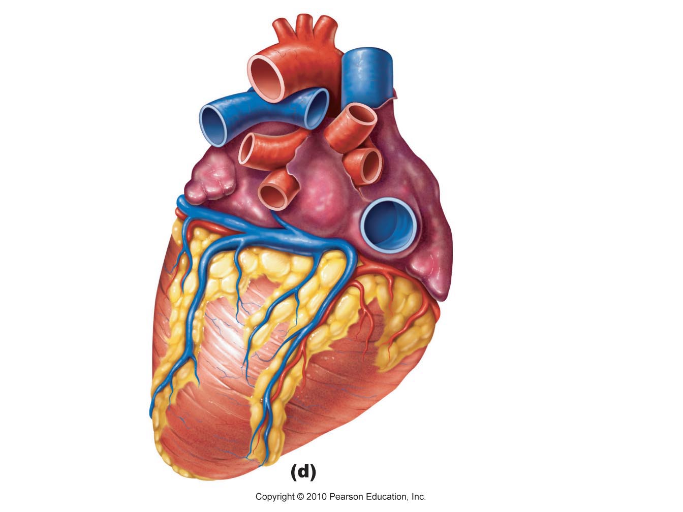
39 structure of the heart without labels
13+ Heart Diagram Templates - Sample, Example, Format Download Color Heart Diagram Sample Format Free Download. cdhb.health.nz This colored heart diagram is a graphic representation of the organ which can be used for presentations and videos about the subject of human heart. The picture is in a coloured format and is available for a free download. Free Download. The Anatomy of the Heart - Quiz 1 - Free Anatomy Quiz The circulatory system - lower body image, with blank labels attached. The circulatory system - a PDF file of the upper and lower body for printing out to use off-line. Describe and explain the function of the circulatory system - The circulatory system consists of the heart, the blood vessels (veins, arteries, and capillaries), and the blood.
Structure of the Heart | Biology for Majors II - Lumen Learning In humans, the heart is about the size of a clenched fist, and it is divided into four chambers: two atria and two ventricles. There is one atrium and one ventricle on the right side and one atrium and one ventricle on the left side. The atria are the chambers that receive blood, and the ventricles are the chambers that pump blood.

Structure of the heart without labels
Human Heart - Anatomy, Functions and Facts about Heart The human heart is about the size of a human fist and is divided into four chambers, namely two ventricles and two atria. The ventricles are the chambers that pump blood and atrium are the chambers that receive blood. Among which both right atrium and ventricle make up the "right heart," and the left atrium and ventricle make up the "left heart." Anatomy of the Heart - Medical Animation - YouTube This medical animation demonstrates the anatomy of the human heart, while explaining how the cardiovascular system functions. Explore more of our medical ani... Basic Anatomy of the Heart - Health Encyclopedia - University of ... The adult human heart is about the size of a fist. The heart beats at an average rate of 80 times a minute. That's about 115,000 times in one day, or about 42 million times in a year. In a 70-year lifetime, an average human heart will beat more than 2.5 billion times. The heart works hard even when you are at rest.
Structure of the heart without labels. Heart Anatomy | Anatomy and Physiology - Lumen Learning The wall of the heart is composed of three layers of unequal thickness. From superficial to deep, these are the epicardium, the myocardium, and the endocardium. The outermost layer of the wall of the heart is also the innermost layer of the pericardium, the epicardium, or the visceral pericardium discussed earlier. Figure 6. Human Heart Diagram Without Labels - Labelling Worksheet The human heart is a muscle made up of four chambers, these are: Two upper chambers - the left atrium and right atrium Two lower chambers - the left and right ventricles. It's also made up of four valves - these are known as the tricuspid, pulmonary, mitral and aortic valves. Heart Anatomy: Labeled Diagram, Structures, Function, and Blood Flow Chambers of the Heart Let's begin with the chambers of the heart. There are 4 chambers, labeled 1-4 on the diagram below. To help simplify things, we can convert the heart into a square. We will then divide that square into 4 different boxes which will represent the 4 chambers of the heart. Figuring Out Cardiac Anatomy: Your Heart - dummies Parietal pericardium: Beyond the pericardial cavity, working your way out to the outside of the heart, this outermost layer of the heart is a thin, white covering made of fibrous connective tissue that joins the major blood vessels (such as the aorta) to the sternum and diaphragm. Your heart is not just floating in your chest.
Structure of the Heart | SEER Training The human heart is a four-chambered muscular organ, shaped and sized roughly like a man's closed fist with two-thirds of the mass to the left of midline. The heart is enclosed in a pericardial sac that is lined with the parietal layers of a serous membrane. The visceral layer of the serous membrane forms the epicardium. Layers of the Heart Wall Heart Diagram with Labels and Detailed Explanation - BYJUS Diagram of Heart. The human heart is the most crucial organ of the human body. It pumps blood from the heart to different parts of the body and back to the heart. The most common heart attack symptoms or warning signs are chest pain, breathlessness, nausea, sweating etc. The diagram of heart is beneficial for Class 10 and 12 and is frequently ... How to Draw the Internal Structure of the Heart (with Pictures) To finish drawing the aorta, draw three nubs at the top of the loop. After you draw these, erase the lines connecting from one side of the bottom of the nub to the other. Add tilted circles to the top of all of the nubs. Draw a circle at the bottom of the aorta, adjacent to the left ventricle. Human Heart Diagram Labeled - Science Trends The heart's atrioventricular valves are structures that join the atria and ventricles of the heart together. This group of valves is comprised of the tricuspid valve and the mitral valve. Beyond this, there is a structure referred to as the aortic valve which separates the left ventricle and the aorta.
Label the heart — Science Learning Hub Label the heart — Science Learning Hub Label the heart Add to collection In this interactive, you can label parts of the human heart. Drag and drop the text labels onto the boxes next to the diagram. Selecting or hovering over a box will highlight each area in the diagram. Pulmonary vein Right atrium Semilunar valve Left ventricle Vena cava Human Heart (Anatomy): Diagram, Function, Chambers, Location in Body The heart is a muscular organ about the size of a fist, located just behind and slightly left of the breastbone. The heart pumps blood through the network of arteries and veins called the... Heart: illustrated anatomy - e-Anatomy - IMAIOS This interactive atlas of human heart anatomy is based on medical illustrations and cadaver photography. The user can show or hide the anatomical labels which provide a useful tool to create illustrations perfectly adapted for teaching. Anatomy of the heart: anatomical illustrations and structures, 3D model and photographs of dissection. Heart Blood Flow | Simple Anatomy Diagram, Cardiac Circulation ... - EZmed Step 2 involves the left atrium, the chamber of the heart that receives oxygenated blood from the lungs via the pulmonary veins. 3. Mitral Valve Step 3 involves the mitral valve. During diastole, when the heart is relaxed and filling with blood, the oxygenated blood from the left atrium will flow to the left ventricle.
Label every structure on the figure of the heart: image | Study.com
Heart Labeling Quiz: How Much You Know About Heart Labeling? Here is a Heart labeling quiz for you. The human heart is a vital organ for every human. The more healthy your heart is, the longer the chances you have of surviving, so you better take care of it. Take the following quiz to know how much you know about your heart. Questions and Answers 1. What is #1? 2. What is #2? 3. What is #3? 4. What is #4?
.png)
Parts Of The Heart - ProProfs Quiz
A Labeled Diagram of the Human Heart You Really Need to See The human heart, comprises four chambers: right atrium, left atrium, right ventricle and left ventricle. The two upper chambers are called the left and the right atria, and the two lower chambers are known as the left and the right ventricles. The two atria and ventricles are separated from each other by a muscle wall called 'septum'.

In this diagram they are showing the function of the heart as they have labels to the parts of ...
Human Heart - Diagram and Anatomy of the Heart - Innerbody The heart is a muscular organ about the size of a closed fist that functions as the body's circulatory pump. It takes in deoxygenated blood through the veins and delivers it to the lungs for oxygenation before pumping it into the various arteries (which provide oxygen and nutrients to body tissues by transporting the blood throughout the body).

Cardiovascular System – The Internet’s Best Anatomy & Physiology Tutorial and Study Resource ...
Heart Drawing With Labels - Anatomy Health Heart Human Science Human ... Internal structure of human heart shows four chambers viz. Draw coronary arteries on the heart as shown. · the arteries carry the blood rich in oxygen from the heart to different parts of the body. Two atria and two ventricles and couple of blood vessels opening into them.
Free Blank Heart Diagram, Download Free Blank Heart Diagram png images, Free ClipArts on Clipart ...
Anatomy of a Human Heart - uofmhealth Located between the lungs in the middle of the chest, the heart pumps blood through the network of arteries and veins known as the cardiovascular system. It pushes blood to the body's organs, tissues and cells. Blood delivers oxygen and nutrients to every cell and removes the carbon dioxide and other waste products made by those cells.

Please label the following heart anatomy:
Cardiovascular System - Human Veins, Arteries, Heart Cardiovascular System Anatomy The Heart. The heart is a muscular pumping organ located medial to the lungs along the body's midline in the thoracic region. The bottom tip of the heart, known as its apex, is turned to the left, so that about 2/3 of the heart is located on the body's left side with the other 1/3 on right.

Heart label diagram
circulatory system worksheet without labels - Google Search | Heart ... Shows a picture of a heart with a description of how blood flows through the heart, focusing on the chambers, vessels, and valves. Students are asked to label the heart and trace the flow of blood. Questions at the end of the activity reinforce important concepts about the heart and circulatory system.
Structure of heart with proper labeling - YouTube
Heart Anatomy: size, location, coverings and layers : Anatomy & Physiology Layers of the Heart Wall The heart wall is composed of three layers: the epicardium, myocardium, and endocardium. Location of the heart in the mediastinum. The superficial epicardium is the visceral layer of the serous pericardium. The middle layer is the myocardium and is composed mainly of cardiac muscle and forms the bulk of the heart.

editorial « Graphic Design, Photorealistic CGI, Information Graphics, Technical Illustration ...
Anatomy of the Human Heart - Physiopedia Anatomy. The heart has a somewhat conical form and is enclosed by the pericardium. It is positioned posteriorly to the body of the sternum with one-third situated on the right and two-thirds on the left of the midline. The heart measures 12 x 8.5 x 6 cm and weighs ~310 g (males) and ~255 g (females) Relations.

Science Stuff.: August 2010
Label Heart Anatomy Diagram Printout - EnchantedLearning.com This cycle is then repeated. Every day, the heart pumps about 2,000 gallons (7,600 liters) of blood, beating about 100,000 times. Label the heart anatomy diagram below using the heart glossary. Note: On the diagram, the right side of the heart appears on the left side of the picture (and vice versa) because you are looking at the heart from the ...
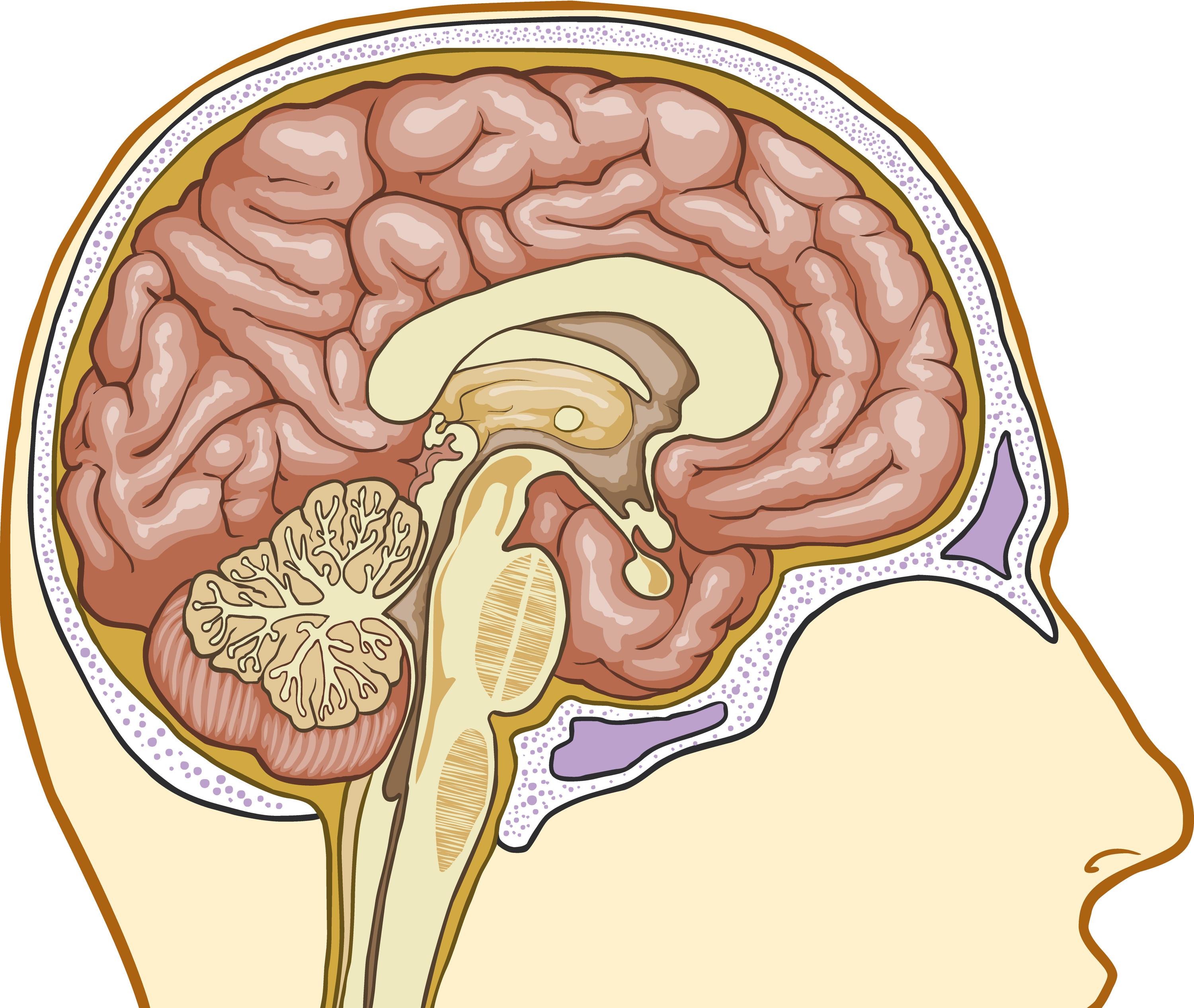
Basic Structure Of The Thalamus - Interactive Biology, with Leslie Samuel
Basic Anatomy of the Heart - Health Encyclopedia - University of ... The adult human heart is about the size of a fist. The heart beats at an average rate of 80 times a minute. That's about 115,000 times in one day, or about 42 million times in a year. In a 70-year lifetime, an average human heart will beat more than 2.5 billion times. The heart works hard even when you are at rest.
.png)
Parts Of The Heart - ProProfs Quiz
Anatomy of the Heart - Medical Animation - YouTube This medical animation demonstrates the anatomy of the human heart, while explaining how the cardiovascular system functions. Explore more of our medical ani...

Long Bone Label The Structure The Long Bone And Labels Label Long Bone Diagram Blank Bone ...
Human Heart - Anatomy, Functions and Facts about Heart The human heart is about the size of a human fist and is divided into four chambers, namely two ventricles and two atria. The ventricles are the chambers that pump blood and atrium are the chambers that receive blood. Among which both right atrium and ventricle make up the "right heart," and the left atrium and ventricle make up the "left heart."

Brain Labeling Diagram - koibana.info | Brain diagram, Basic anatomy and physiology, Human brain ...

Heart Anatomy Quizzes and Flashcards – sciencemusicvideos

Heart Diagram Labeled
Post a Comment for "39 structure of the heart without labels"